Research / Curved planar reformatting
Following flow across the dissection flap.
Flap-referenced curved planar reformation (CPR) straightens the aorta and separates flow into and out of the false lumen, for inspection of small flap-crossing jets throughout the cardiac cycle.
Submitted to JCMR
Flap-Referenced Curved Planar 4D Flow MRI for Tear Visualization in Aortic Dissection
Sebastian Cohn, Elizabeth Weiss, Shaunak Roy, Michael Markl, and Bradley D. Allen
My contribution: method development with collaborators, data analysis, and manuscript drafting.
The visualization
Unfold the anatomy. Separate the flow.
The interface between the true and false lumens guides the straightened view. A direction derived from the lumen centerlines separates flow toward each lumen.
The contribution combines an anatomy-referenced display with directional flow, so an observer can inspect where blood crosses the flap throughout the cardiac cycle.
The imaging problem
A projection shows speed. The flap gives it context.
In aortic dissection, tears allow blood to pass between the true and false lumens. Small crossing jets can be obscured by overlapping anatomy, and velocity magnitude alone does not retain flow direction relative to the flap.
How the flap-referenced view is constructed
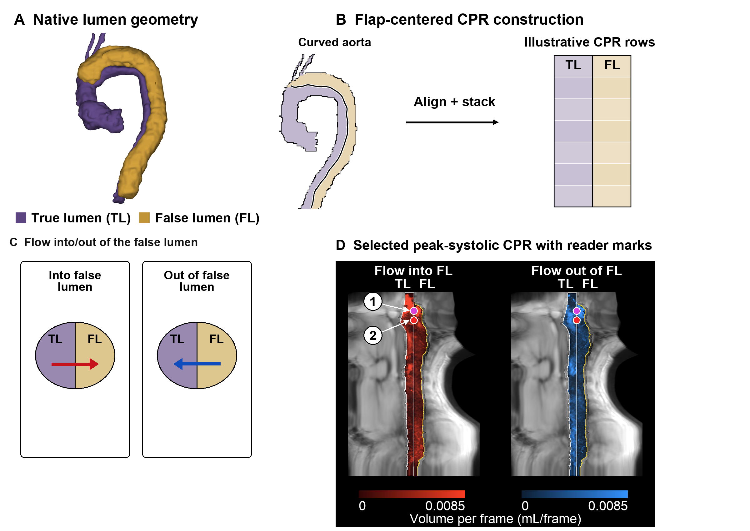
The lumen interface guides the straightening path. A direction derived from the lumen centerlines separates flow toward each lumen; projected velocity is converted to a voxelwise signal in mL/frame.
Open full-size figure (new tab)01
Find a path along the flap
Using the true- and false-lumen masks, identify false-lumen voxels next to the true lumen. Move false-lumen centerline points toward this interface and smooth the resulting path.
02
Unfold the curved anatomy
Sample along the path and stack the planes into a straightened view. This brings a longer extent of the dissection flap into one display.
03
Separate the flow directions
Project velocity along the direction between lumens and express it as volume per frame at each voxel. Show flow into and out of the false lumen in separate, time-resolved panels.
Evaluation
A paired reader study in 36 participants.
Two physician readers compared CPR with sagittal velocity-magnitude maximum intensity projection (MIP), one scan per participant. A reader-marked tear required a discrete jet crossing the flap at one location for at least three consecutive frames. All analyses excluded aortic zone 0.
| Reader | CPR | MIP | Adjusted p |
|---|---|---|---|
| Reader 1 | 52 | 35 | 0.025 |
| Reader 2 | 54 | 47 | 0.674 |
Two-sided Wilcoxon signed-rank tests paired by scan; Holm adjustment across all eight count comparisons in the submitted manuscript.
What the comparison supports
Both readers marked more tears with CPR in total; the paired difference was significant only for Reader 1. CPR marks occurred in more aortic zones containing a CTA/MRA tear, but also in more zones without a recorded reference tear.
Counts and shared zones do not confirm that individual marks correspond to the same tears. These results do not establish diagnostic superiority.
Limitations and next steps
From clearer displays to confirmed findings.
What remains unresolved
This single-center study included 36 participants and two readers. CPR changed both geometry and signal, so the comparison cannot isolate the contribution of straightening alone. Display type was apparent, and no separate washout session was used.
One reader provided the CTA/MRA reference. Comparisons used counts and zones rather than matching individual tears, and the reference scan could be from a different time point. Review time and clinical utility remain to be evaluated.
Connecting visualization and measurement
The next evaluation should match individual reader marks to reference tears, including locations marked with only one display. A projection using the same directional flow signal would help separate the effects of geometry and signal content.
This visualization work complements the tear detector: one supports human inspection of flow exchange; the other develops automated, structured measurements from 4D Flow MRI.
Explore the tear detectorThis work is submitted to the Journal of Cardiovascular Magnetic Resonance and is not yet published. Participant-level clinical imaging data are not publicly available; analysis and display-generation code are available from the corresponding author on reasonable request.